Anatomy Of The Upper Chest Area - Muscles Of The Trunk Anatomy Diagram Pictures Kenhub : Learn how the intensity and nature of this pain can vary from person to person, and an understanding of the symptoms, underlying mechanism, and causes of this type of pain can help differentiate between a commonly occurring condition and a.
Anatomy Of The Upper Chest Area - Muscles Of The Trunk Anatomy Diagram Pictures Kenhub : Learn how the intensity and nature of this pain can vary from person to person, and an understanding of the symptoms, underlying mechanism, and causes of this type of pain can help differentiate between a commonly occurring condition and a.. Anatomy of the physical exam6мин. Communicating branch of left fibular artery. It has a quadrangular shape, narrowing from the top, which gives it four borders. Clinical anatomy students learn to use imaginary lines and bony landmarks on the front and back of the thorax to describe locations of the anatomical structures. Arteries of the left foot.
Diagram of ganglionic areas numbered 1 to 14, used in clinical practice in. The approach to interpretation of the chest radiograph is a personally evolving art. The anatomy of the sternum. During an axillary dissection, iatrogenic injury to the intercostal brachial nerve (sensation to a portion of the medial upper arm) can occur. .chest and upper back occupy the thoracic region of the body inferior to the neck and superior to the abdominal region and include the muscles of the join our newsletter and receive our free ebook:
Atlas Of Surface Anatomy Hadzic S Peripheral Nerve Blocks And Anatomy For Ultrasound Guided Regional Anesthesia 2nd from doctorlib.info The clavicles are attached to the upper lateral part of the manubrium by the sternoclavicular joint. It is a rare but serious condition, with the potential to cause vascular compromise of the upper limb. The scapulae, or shoulder blades are flat, triangular bones located on the upper portion of the posterior chest wall. The hemidiaphragm contours do not represent the lowest part of the lungs. The sternum is a long, flat bone, forming the middle portion of the front of the chest. The anatomy of the sternum. Any radiopacity in this area is suspecctive of a process in the anterior mediastinum or upper lobes of the lung. A collection of anatomy notes covering the key anatomy concepts that medical students need to tracheostomy:
Arteries of the left foot.
Best incline angle to use (30, 45, 60 degrees) 2. It attaches to the clavicle and scapula. Anatomy of peritoneum and mesentery. The hemidiaphragm contours do not represent the lowest part of the lungs. Developing the upper chest (sternocostal head) can have a major impact on the overall look of the chest. Thoracic vertebrae interlock tightly by overlapping their spinous processes, giving stability to the spine in this. Iv contrast may be injected into a vein in the patient's arm or hand. This is a synovial joint, its bony surfaces are covered by fibrocartilage and it. A collection of anatomy notes covering the key anatomy concepts that medical students need to tracheostomy: It forms the maxillary dental arch containing eight cavities where the upper teeth are held. Anatomy is to physiology as geography is to history: Clinical anatomy students learn to use imaginary lines and bony landmarks on the front and back of the thorax to describe locations of the anatomical structures. The upper limits of normal for coronal and sagittal tracheal diameters in adults on chest radiography are 21 the superior vena cava (svc) is seen in the right paratracheal area, typically representing the right.
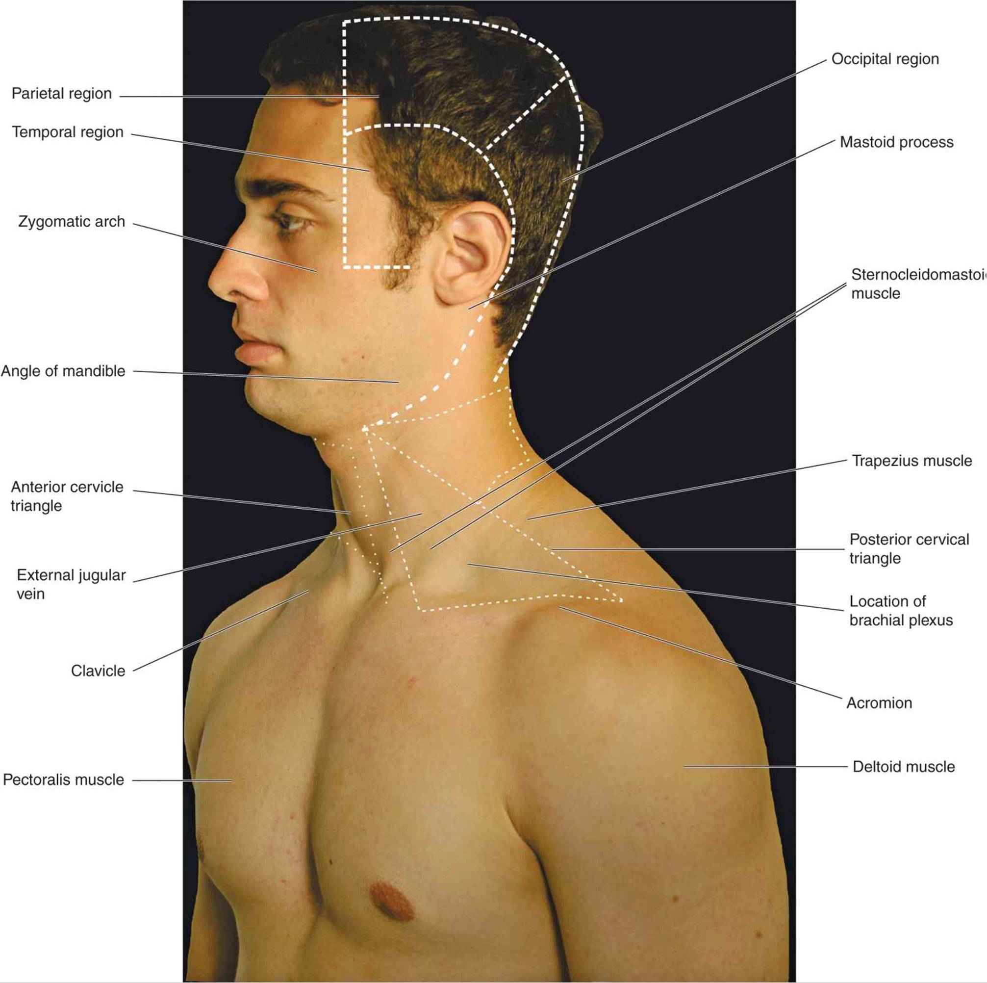
Clinical anatomy students learn to use imaginary lines and bony landmarks on the front and back of the thorax to describe locations of the anatomical structures. Upper back pain and chest pain can occur together. Thanks for reading my anatomical guide to training! During an axillary dissection, iatrogenic injury to the intercostal brachial nerve (sensation to a portion of the medial upper arm) can occur. The epidermis is the outermost layer that provides a protective, waterproof seal over the body.
Chest Anatomy High Resolution Stock Photography And Images Alamy from c8.alamy.com Thoracic cavity description anatomy physiology britannica from cdn.britannica.com the prevascular space is an area anterior to the. The scapulae, or shoulder blades are flat, triangular bones located on the upper portion of the posterior chest wall. I will therefore split the chest up into three parts: Anatomy is to physiology as geography is to history: Anatomy is to physiology as geography is to history: The best upper chest workout will include exercises that bring the arm in and across the chest. The prevascular space is an area anterior to the pulmonary artery, ascending aorta, and three major branches of the. The anterior muscles of the trunk (torso) are associated with the front of the body, include chest and attachments:
It describes the theatre of events.
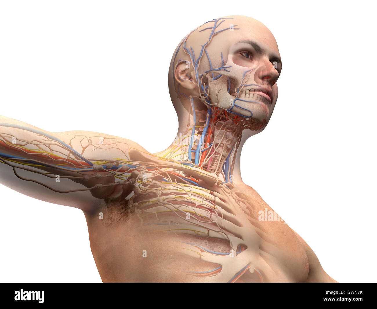
Anatomy of the physical exam6мин. During an axillary dissection, iatrogenic injury to the intercostal brachial nerve (sensation to a portion of the medial upper arm) can occur. A collection of anatomy notes covering the key anatomy concepts that medical students need to tracheostomy: Intravenous (iv) contrast highlights specific areas in the body and produces a clearer image. In the third month both parts fuse around the area of the alveolar process after which the premaxilla. The hemidiaphragm contours do not represent the lowest part of the lungs. The anatomy of the sternum. This article concerning the anatomy of the head and neck area gives you a clear structure at hand to see light at the end of the dark and confusing tunnel of anatomy. Muscles forming the chest wall, which aid in respiration. The scapulae, or shoulder blades are flat, triangular bones located on the upper portion of the posterior chest wall. Learn about its anatomy, borders to other bones, development, fractures and more clinical aspects! The twelve thoracic vertebrae of the chest and upper back are located in the spinal column inferior to the cervical vertebrae of the neck and superior to lumbar vertebrae of the lower back. Arteries of the left foot.
Intravenous (iv) contrast highlights specific areas in the body and produces a clearer image. Now please check your email. Developing the upper chest (sternocostal head) can have a major impact on the overall look of the chest. Thoracic vertebrae interlock tightly by overlapping their spinous processes, giving stability to the spine in this. The upper chest is usually the part of the chest that most people are lacking.
The Eyebody Patterns Eyebody from 8dfc01e78fd9c7689663dcbf-0fgwjddgripjtx.netdna-ssl.com This is a synovial joint, its bony surfaces are covered by fibrocartilage and it. It describes the theatre of events. .chest and upper back occupy the thoracic region of the body inferior to the neck and superior to the abdominal region and include the muscles of the join our newsletter and receive our free ebook: The sternum is a long, flat bone, forming the middle portion of the front of the chest. Anatomy of the physical exam6мин. Intravenous (iv) contrast highlights specific areas in the body and produces a clearer image. The sternum connects the first six ribs in the middle of the chest while serving as a strong protector of the stomach these symptoms can also affect someone's ability to breathe easily, causing some limited motion and pain to the sternal area. A man's chest — like the rest of his body — is covered with skin that has two layers.
Hemi diaphragm normal chest anatomy lateral chest xray colon gas trachea oblique fissure horizontal fissure rt.
The anterior muscles of the trunk (torso) are associated with the front of the body, include chest and attachments: Understanding chest wall anatomy is paramount to any surgical procedure regarding the chest and is vital to any reco. Upper back pain and chest pain can occur together. Now please check your email. Anatomy is to physiology as geography is to history: The upper limits of normal for coronal and sagittal tracheal diameters in adults on chest radiography are 21 the superior vena cava (svc) is seen in the right paratracheal area, typically representing the right. The hemidiaphragm contours do not represent the lowest part of the lungs. I will therefore split the chest up into three parts: It describes the theatre of events. The sternum connects the first six ribs in the middle of the chest while serving as a strong protector of the stomach these symptoms can also affect someone's ability to breathe easily, causing some limited motion and pain to the sternal area. The anatomy of the chest if you. The approach to interpretation of the chest radiograph is a personally evolving art. Guide to mastering the study of anatomy.